Can you introduce us to the main materials studied with X-ray tomography?
EPFL carries out research in many fields. Thus, we regularly study, within our PIXE platform, materials such as wood, concrete, rocks as well as composite materials. We also have the opportunity to scan insects, archaeological pieces, bones as well as mechanical parts.
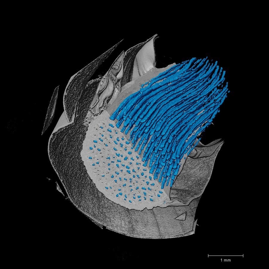
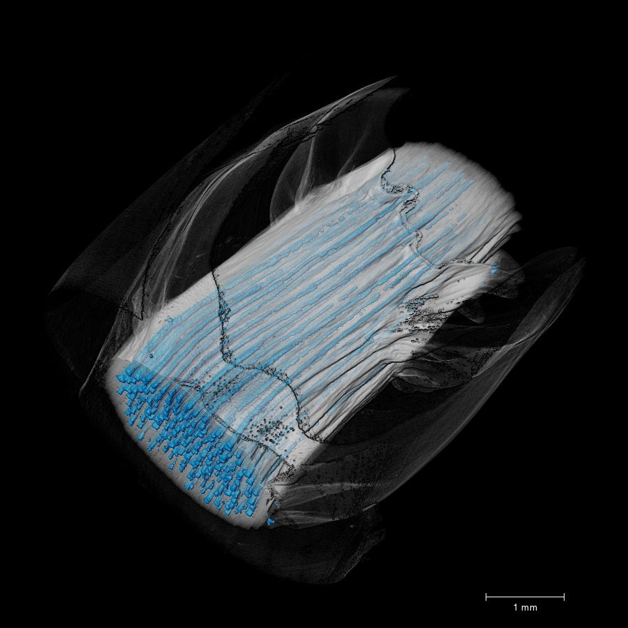
Rhizome wood in 3D
What is the main interest for EPFL to use X-Ray tomography ?
EPFL is a renowned school that trains engineers, especially in civil and materials engineering. X-ray CT allows us to characterize, with great precision, the different materials studied and developed here.
The versatility of the UltraTom, allows us to develop different experiment grounds to perform in-situ inspections and measurements. We currently have two units for tensile and compression tests. In one of them, we can also control humidity. We also have a unit for shear tests and plan to develop others in the near future thanks to UltraTom testing capabilities.
For which applications is X-Ray CT most effective ?
We are using X-ray CT more and more for in-situ testing. A laboratory of the EPFL, the CCLAB, regularly mandates us to perform in-situ tests. For example, in the images below (carousel of glassfiber images), we see a section of a composite material during a tensile test. This test allows us to see the deformation mechanics as well as the crack initiations in the material up to its breaking point, over time.
In-situ testing on a glassfiber With UltraTom - cracks over time
What are the benefits of X-ray ct for material analyses?
The main benefits for us at EPFL are; the non-destructive aspect, the 3D visualization of the outside and inside of the sample as well as the possibility to do in-situ tests (4D inspection over time). Indeed, with X-ray CT, it is not necessary to prepare the sample, to cut it and to polish it like for a microscope analysis for example. It's easier and faster for us. In addition, X-ray CT gives us a complete mapping of the structure and defects of the sample.