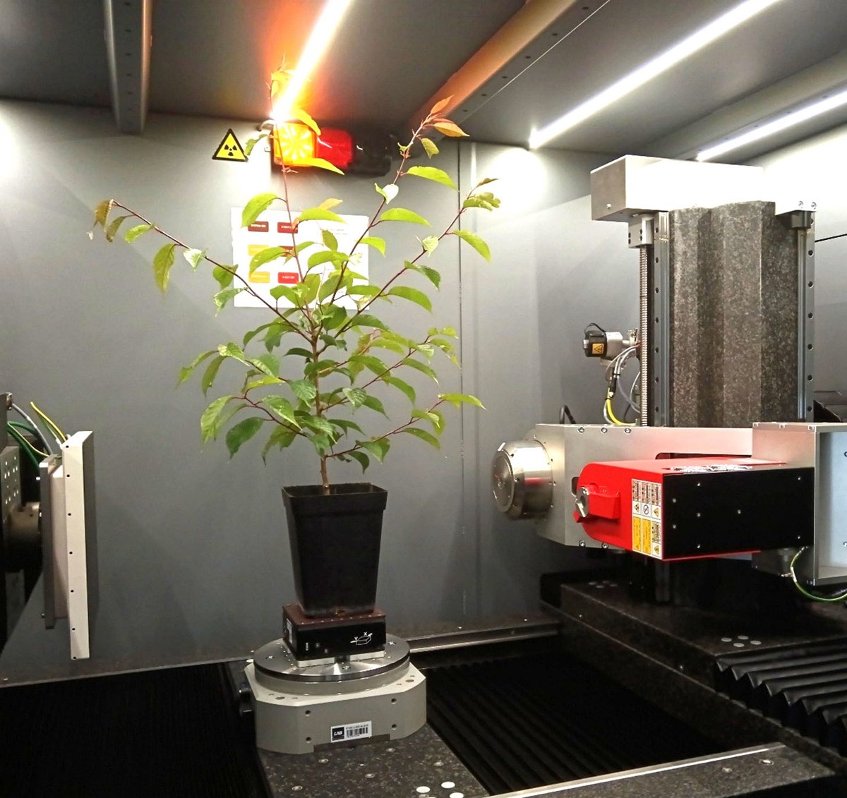
Small potted tree inside the EasyTom XL cabinet (photo: E. Badel)
How does a tree die of drought?
Answering that question means looking well beyond the withered leaves or dead branches visible from the outside.
At the PIAF laboratory, Éric Badel and his colleagues study how trees function and acclimate to their environment, in particular to drought and wind. Their research focuses on tree mechanics and hydraulics, two fields that are especially important for understanding the effects of climate change.
"To see what happens inside the wood, X-ray microtomography has become an invaluable tool. It makes it possible to explore the tree in 3D, right at the heart of its structure, without cutting the sample." explains Eric.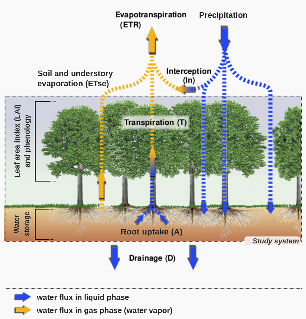
Water flow in trees ©INRAE UMR Silva
When a tree’s hydraulic system fails
In a tree, water is taken up by the roots and carried to the leaves through a network of vessels. This movement is driven by transpiration: at the leaf surface, water evaporates. Transpiration thus acts as a pump, drawing water up through the tree from the roots to the leaves. As a result, the water column inside the tree is held under tension.
During a drought, this water flow can be disrupted. When the water available in the soil is no longer sufficient while the leaves keep transpiring, the water columns can break. Vessels empty of water and fill with air. This is known as vascular embolism. Affected vessels can no longer transport water normally.Healthy vascular network vs. vascular embolism. Case of a poplar.
Good health
3D X-ray micro-tomography view of the vascular network of a poplar (photo: P. Conchon)
×Drought impact
Cross-section of a drought-stressed poplar stem. Embolized vessels fill with air (black dots) and can no longer carry raw sap from the roots to the leaves. (Photo: E. Badel)
×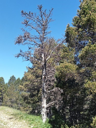
Tree killed by severe water stress (summer 2022)
Embolism can kill a tree
When embolism becomes widespread, it can be lethal for the tree. Embolism in a tree’s vessels is irreversible.
“If only a few vessels are affected, the tree can still recover. But when embolism reaches a very large share of the vascular network, it can lead to the death of the tree,” explains Éric Badel.
“Water also acts as a coolant,” Éric continues. “During droughts, another consequence of the water flow stopping is that leaves are no longer cooled and can be scorched by the heat.”
This internal embolism is precisely what micro-CT can observe directly. It provides three-dimensional, localized, non-destructive information, making it possible in particular to tell water-filled vessels from air-filled ones and thereby to assess the level of embolism in the tissues observed.Observing without causing the very phenomenon you are trying to measure
The contribution of X-ray microtomography is not limited to image quality, however. It has also helped call some experimental results into question.
Éric explains that, before tissues could be observed directly, the study of embolism relied in particular on other methods that required cutting out sections of branches.
The difficulty arises with certain species whose vessels are exceptionally long. Oak, which Éric mentions, is one of them: when a vessel can exceed one meter in length and the sample taken is only 30 cm long, the cut opens the vessel from end to end. It then empties, regardless of the drought the tree has actually experienced. This led to the publication of many “odd” curves that were in fact the product of this measurement artifact.
In other words, preparing the sample can trigger the very event the experiment is meant to measure.
Spruce vascular network
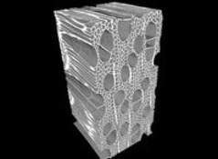
Poplar vascular network
Here, then, micro-CT does not simply provide a more detailed image: it has also made it possible to validate scientific hypotheses and to check the reliability of other experimental methods.
3D observation of the network of vessels that conducts sap, in a spruce (left) and a poplar (right).
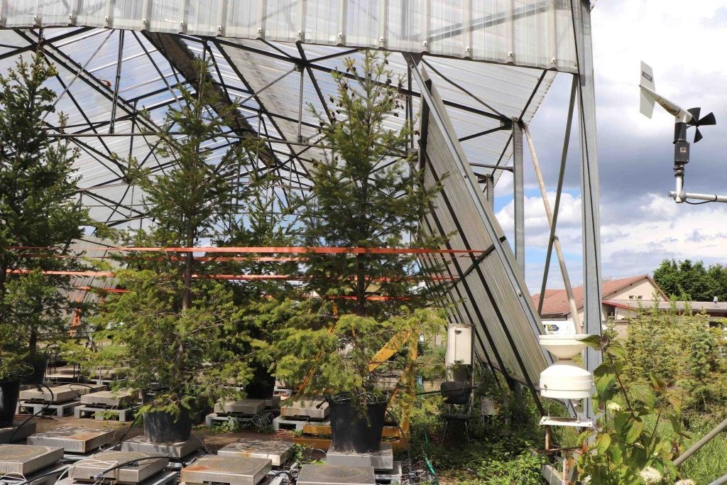
Studying fir trees under severe drought at PIAF @MP-Demarty
From the synchrotron to the lab: following the same tree over time
Another challenge is monitoring change over time. Once a branch has been cut, its condition can be observed in great detail, but the biological process is interrupted. The laboratory therefore wanted to carry out in vivo observations and follow a single organism over time.
To do this, the researchers turned to synchrotrons, first in Grenoble and then notably at SOLEIL, near Paris. But this approach could not offer the same availability as equipment housed directly in the laboratory.
Having the new CT scanner on site now makes it possible to carry out these in vivo observations and to monitor the same sample over successive scans. Éric Badel mentions in particular the monitoring of different tree species subjected to drought conditions, with scans taken at different points in time to observe their response, with the aim of selecting the most resistant species.
This changes the nature of the experiment: rather than comparing several individuals sacrificed at different stages, researchers can now follow the evolution of a single organism.Plants: a particularly delicate object to scan
This in vivo observation nonetheless raises a fundamental difficulty. Tomography does let you observe a sample without cutting or mechanically altering it. But exposure to X-rays is not necessarily neutral for a living organism.
"The radiation is ionizing. When a living plant is monitored over time, the dose it receives becomes an experimental variable in its own right. Part of the know-how developed at PIAF therefore lies in adapting the acquisition settings and the way samples are prepared and housed to these biological materials, so as to limit any change in them during observation." explains Eric.
For example, Éric and his teams seek to reduce the dose, sometimes at the cost of a compromise on spatial resolution or image quality. The goal is to obtain the information needed while allowing the plant to keep growing.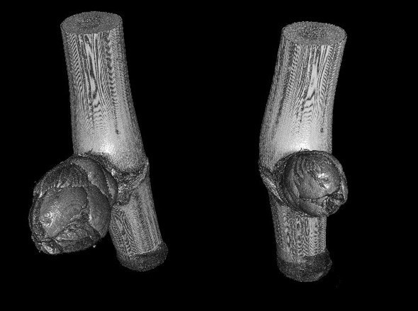
CT Monitoring the development of a bud (photo: P. Conchon)
An experiment on a bud illustrates this approach. The team subjected it to several successive scans, about one week apart, to check whether it kept developing after being scanned. The bud did continue to grow. The aim was not to draw an immediate, general biological conclusion, but to explore acquisition conditions compatible with monitoring living material.
The researchers are therefore still in an exploratory phase as they work out the right conditions for living plants.
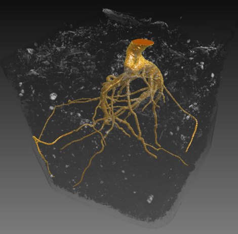
Micro-CT observation of a poplar root system in a pot (photo: P. Conchon)
Observing roots without digging them up
This ability to observe without destroying the plant under study is opening up other research paths.
Éric tells me that a PIAF researcher is working on root systems. The first tomography experiments aim to determine what 3D imaging can bring to this field.
One of the questions raised concerns how the root system is distributed in the soil depending on growing conditions.
“Conventional observation methods bring an obvious difficulty here: pulling roots out of the soil lets you study them, but it removes their support, can change their configuration and ends the experiment,” Éric points out. “Tomography, by contrast, lets you observe the 3D organization of roots in their environment, and how it changes over time, without having to dig them up.”
This research is still at the stage of initial experiments in the laboratory.Tomography, now a reference method
What was exceptional only a few years ago is now part of PIAF’s research protocols.
For work on embolism, Éric Badel explains that the laboratory now has tomography protocols that are used on a fairly routine basis.
“Tomography has become a ‘standard’ method. When a PhD project deals with embolism, tomography campaigns can be built into the experimental work. If there is any doubt about the level of embolism obtained with another method, tomography has become, for the team, a reliable way to get direct access to observation. It has become a reference method.”
This change is significant: according to Éric, this systematic use of tomography did not exist ten years ago.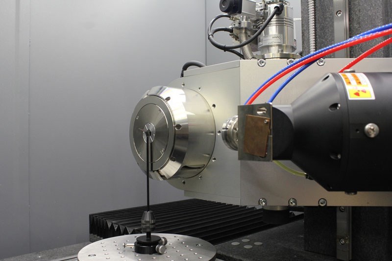
The EasyTom XL’s dual tube
Nano-CT: going down another scale
The next frontier is already taking shape.
The EasyTom XL has two X-ray sources, allowing the team to carry out both micro-CT and nano-CT. With a nano source, the team now aims to go down in scale, moving from observing tissues or cells to structures close to the cell wall of the wood.
Another avenue is the observation of plant samples in a frozen state. Researchers in the laboratory work on winter biology and tree frost resistance. Being able to keep a sample frozen during a tomographic acquisition would make it possible to explore these phenomena under new conditions. Éric Badel says he wants to explore this further with his biologist colleagues, notably by developing systems suited to housing such samples.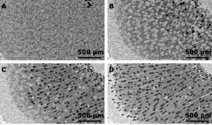
Time-lapse monitoring of a poplar sample under drought © INRAE
Seeing without destroying,
but above all seeing differently
From embolism to bud growth, from wind-stressed roots to biobased materials, the applications may seem very far apart.
Yet they all rest on the same experimental question: how can you observe an internal structure without altering the object to the point of changing the phenomenon you are trying to understand?
At the PIAF laboratory, tomography is no longer seen as an exceptional technology to be called on only occasionally. It has gradually become one tool among others at the researchers’ disposal, used when 3D observation, access to the inside of an object or monitoring over time provides information that other methods cannot.
And that may be where its most interesting scientific contribution lies: not simply seeing better, but making observable phenomena that could not be observed without modifying the object under study.
Many thanks to Éric for the time he gave to this conversation about his research and tomography.